Skip to product information
1 of 1
New Year Badge
🏷️ Save up to 70% on all books, discount auto applied

Difiore’s Atlas of Histology with Functional Correlations 12th Edition

Difiore’s Atlas of Histology with Functional Correlations 12th Edition

Regular price $35.00 USD
Regular price $145.15 USD Sale price $35.00 USD
Sale Sold out
Girl in a jacket

Instant Download

Your unique download link appears right after you complete your purchase, and is also sent instantly to the email address you provided.

ISBN

978-1451175615

Authors

Victor P Eroschenko

Edition Number

12th

Copyright Year

January 1, 2012

File Size

77 MB

Publisher

Lippincott Williams & Wilkins

Pages

560

diFiore’s Atlas of Histology with Functional Correlations

diFiore’s Atlas of Histology with Functional Correlations by Victor P. Eroschenko presents human histology through detailed, full-color illustrations and corresponding photomicrographs. It connects the microscopic structure of cells, tissues, and organs with their essential functions, helping students develop a clear understanding of histological organization. Updated content includes an introduction to histological techniques and staining, a cell biology chapter, additional micrographs, and interactive learning resources for study and review.

Key Features
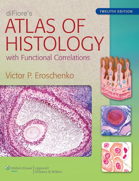
  • Detailed Visual Atlas: Combines realistic, full-color illustrations with actual photomicrographs of histological structures.
  • Structure–Function Correlations: Explains how the microscopic features of cells and tissues relate to their major functions.
  • Cell Biology Coverage: Includes a dedicated chapter introducing cell biology and important cellular processes.
  • Mitosis Illustrations: Presents drawings and representative photomicrographs showing stages of the cell cycle during mitosis.
  • Histological Techniques: Introduces basic histology methods, microscopy, and tissue preparation.
  • Staining Methods: Provides information on common histological stains encountered in laboratory courses.
  • Expanded Image Collection: Includes additional light and transmission electron micrographs for detailed structural examination.
  • Logical Organization: Arranges content into four parts, progressing from methods and microscopy to tissues and organ systems.
  • Interactive Study Resources: Supports learning with an online question bank and an interactive atlas featuring additional micrographs.
  • Exam and Course Preparation: Helps medical and graduate students review histological structures, understand their functions, and reinforce essential concepts.
View full details